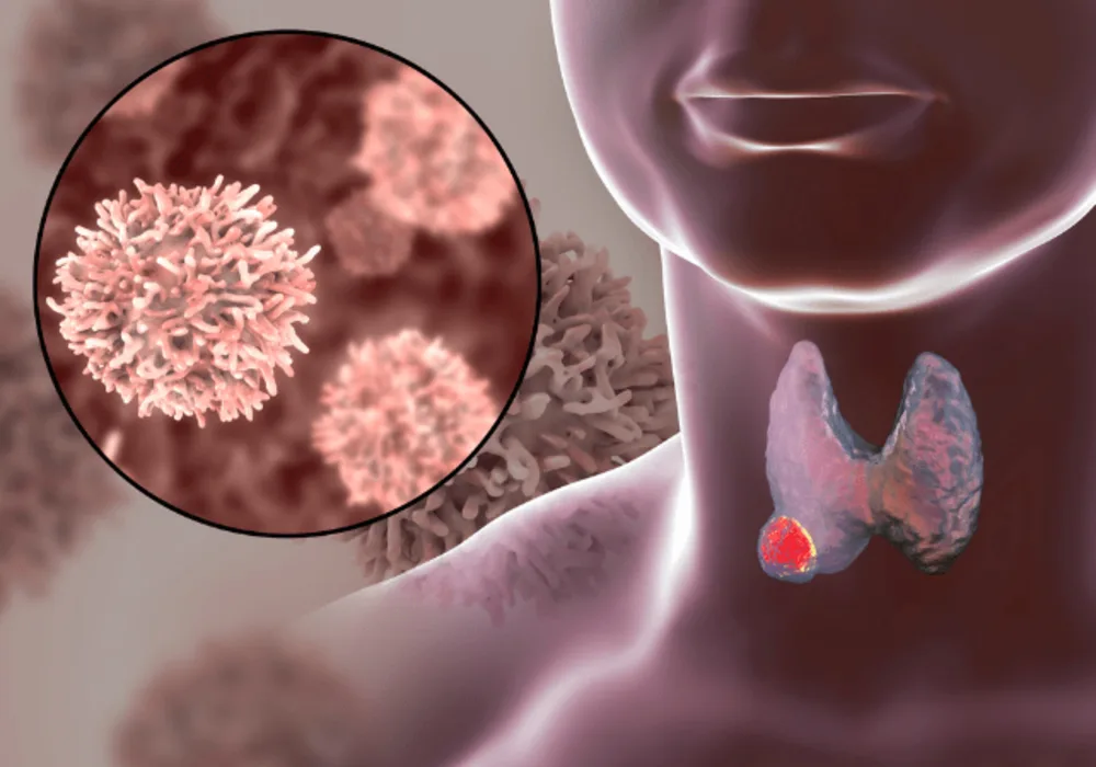
The incidence of thyroid nodules has risen significantly in recent decades, with detection rates as high as 65% in the general population. Despite this, only a small percentage of nodules are cancerous. It's crucial to accurately distinguish between benign and malignant nodules for proper clinical management. While fine-needle aspiration biopsy is the gold standard, it can be invasive and prone to misdiagnosis. Ultrasound is commonly used for non-invasive characterisation, but CT scans are increasingly detecting nodules incidentally. However, management practices for CT-detected nodules vary widely. Morphologic features on CT scans can aid characterisation, but they have limitations in distinguishing between benign and malignant nodules. Radiomics, a method that extracts high-throughput features from images, shows promise in identifying malignancy-associated characteristics not easily discernible by human readers. Previous studies have demonstrated the potential of CT-based radiomics models for differentiating various types of thyroid nodules, with reported high accuracy. However, these studies often lack external validation and have small sample sizes. A recent study published in the American Journal of Roentgenology aims to address these limitations by developing and validating a CT-based radiomics model specifically for distinguishing between benign and malignant thyroid nodules.
Multi-Centre Retrospective Study of CT Examinations Before Surgical Resection
In this retrospective study, two medical centres, Shantou Central Hospital (centre 1) and Sun Yat-Sen Memorial Hospital, Sun Yat-Sen University (centre 2), obtained ethics committee approval. They conducted searches in their electronic medical records (EMR) for patients who underwent preoperative multiphase neck CT scans within two weeks before surgical resection of a thyroid nodule. The search at Centre 1 covered January 2018 to December 2022 and identified 1082 patients, while Center 2's search from January 2020 to December 2022 yielded 710 patients. After excluding patients for various reasons, such as prior thyroid biopsy or the presence of nonthyroid malignancy and removing cases with unclear pathologic diagnoses or CT artefacts, a total of 378 patients (408 nodules) were included in the study. Among these, 278 patients (293 nodules) were from centre 1, with 100 patients (115 nodules) from centre 2. The patients' mean age was 46.3 years, with 86 men and 292 women. Of the nodules, 145 were benign, and 263 were malignant. To create and validate the radiomics model, nodules from centre 1 were randomly divided into training (n = 206) and internal validation (n = 87) sets at a 7:3 ratio. Patients did not overlap between these two sets. All nodules from centre 2 served as the external validation set.
The CT examinations were performed as part of preoperative planning before surgical resection. Each centre used one of two CT scanners. The scans included non-contrast, arterial, and venous phase acquisitions, covering the region from the skull base to the subclavian region. At centre 1, contrast agent injection (Iopromide 300 mgI/mL) rates were 3.0 mL/s, with arterial and venous phase acquisitions initiated 25 seconds and 60 seconds post-injection, respectively. At centre 2, injection of contrast agent (Iopamidol 370mgI/mL) occurred at a rate of 3.0 mL/s, with arterial-phase acquisition starting 4 seconds after reaching 100 HU attenuation in the internal carotid artery, followed by venous-phase acquisition 15 seconds later.
Combined CT-Based Radiomics Model from Noncontrast, Arterial, and Venous Phases
This study aimed to assess the effectiveness of CT-based radiomics models in characterising thyroid nodules. A combined model was developed, incorporating clinical features, morphologic CT features, and radiomic CT features from noncontrast, arterial, and venous phases. This combined model showed a significantly higher AUC (Area Under the Curve) of 0.923 in the external validation set compared to other models. Calibration curve assessment and decision-curve analysis further supported the superiority of the combined model as a diagnostic tool. These results suggest a potential role for radiomics analysis in characterising thyroid nodules detected on CT scans.
The conventional model, which included features such as age, cystic change, edge interruption sign, and abnormal cervical lymph nodes, also performed well with an AUC of 0.827 in the external validation set. Cystic change, a common feature in benign nodules, and the edge interruption sign, indicating possible malignancy, contributed to the model's effectiveness. This study performed better than previous studies focusing solely on morphologic CT features.
Insights and Limitations from Multiphase Imaging in Radiomics Analysis
CT-based radiomics has been explored extensively for differentiating benign and malignant lesions in various organs, including the thyroid. Malignant nodules exhibit distinct radiomic features related to abnormal angiogenesis and changes in cell permeability. Incorporating features from non-contrast, arterial, and venous-phase CT scans in the combined model highlights the importance of multiphase imaging in radiomics analysis for thyroid nodules.
Despite the promising results, the study has limitations, including its retrospective nature, selection bias due to inclusion of only surgically resected nodules, and manual image segmentation introducing variability. Additionally, the study did not compare radiomics models with other imaging modalities such as ultrasound or PET scans.
Nevertheless, the study concludes that the combined model, validated externally, presents a robust method for differentiating benign and malignant thyroid nodules using CT-based radiomics.
Source: American Journal of Roentgenology
Image Credit: iStock


