


Over the past decade, late gadolinium-enhanced (LGE) cardiac MRI has been crucial in assessing left atrial (LA) fibrosis in atrial fibrillation (AF) patients, aiding in predicting AF recurrence post-ablation. This fibrotic burden can guide treatment decisions. Typically, imaging protocols involve a time-consuming diaphragm-navigated (dNAV) sequence. A newer image-navigated (iNAV) approach promises more efficient and predictable scanning, potentially improving clinical implementation. However, the comparative effectiveness of iNAV versus dNAV in visualising LA fibrosis remains unclear. A recent study published in Radiology: Cardiothoracic Imaging aimed to evaluate both qualitatively and quantitatively in individuals with AF.
Study Methodology and Participant Details
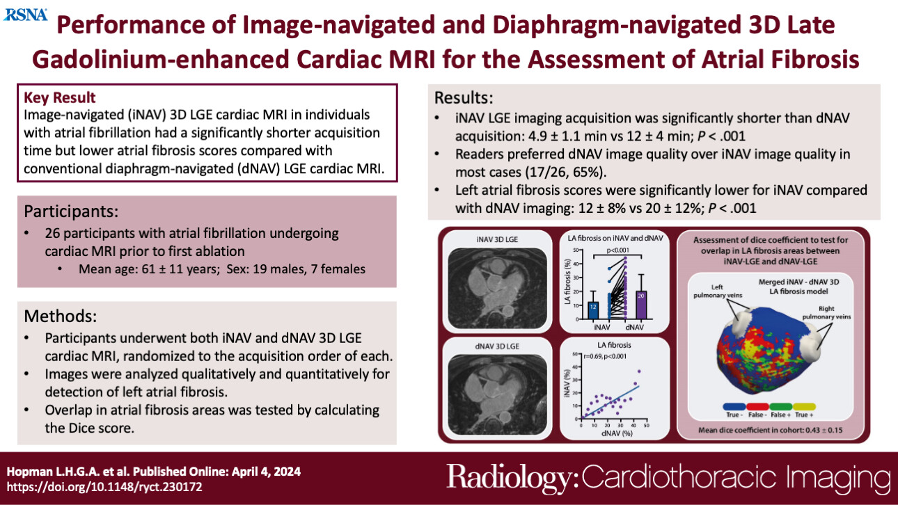
This prospective single-centre study, conducted between April and September 2022, involved participants undergoing cardiac MRI before their first ablation for atrial fibrillation (AF). The study, approved by the Amsterdam University Medical Center Medical Ethics Committee, included 26 consecutive AF participants. It built upon prior research by Munoz et al., comparing iNAV and dNAV 3D LGE techniques in 18 AF participants, focusing on image quality and scan duration. The primary aim was to quantify and localise LA fibrosis using both techniques. Participants, aged over 18, underwent prepulmonary vein isolation cardiac MRI scans, excluding those with general cardiac MRI contraindications, contraindications for gadolinium-based contrast agents, cardiac implantable electronic devices, and mechanical heart valves. Images were acquired using a 1.5-T MRI system and a specific scan protocol. Twenty-six participants (mean age 61 years ± 11 [SD]; 19 [73%] men, seven [27%] women) were included, with most in sinus rhythm during the scan. The time between contrast agent application and scan start differed slightly between iNAV and dNAV sequences.
Impact of Respiratory and Rhythmic Factors on Technique Performance
This study compared two cardiac MRI techniques, iNAV and dNAV, specifically focusing on their efficacy in imaging atrial fibrosis in individuals with atrial fibrillation (AF). The iNAV technique demonstrated notable advantages over dNAV, particularly in terms of acquisition time. On average, iNAV scans took less than 5 minutes, a significant improvement compared to the approximately 12 minutes required for dNAV scans. Moreover, iNAV exhibited a more predictable acquisition time range (3 to 8 minutes) compared to the broader range of 7 to 26 minutes observed with dNAV. While both techniques yielded similar final image quality, dNAV was generally preferred by practitioners due to perceived advantages in clarity and sharpness. However, iNAV showed a distinct advantage in terms of lower fibrotic burden in the left atrium (LA). This finding suggests potential differences in the sensitivity of the two techniques to detect atrial fibrosis. Additionally, there was only modest spatial overlap between fibrosis areas detected by iNAV and dNAV, indicating variability in fibrosis localisation between the two methods.
Implications for Patient Categorisation and Treatment Decisions
Several factors influenced the performance of each technique. Respiratory pattern had a significant impact on dNAV scan quality and duration, whereas iNAV was less affected by irregular respiratory patterns. However, irregularities in heart rhythm, particularly in AF, influenced both techniques similarly, potentially leading to lower scan quality but without a corresponding increase in acquisition time for iNAV. The differences in fibrosis measurement between iNAV and dNAV could have implications for patient categorisation and treatment decisions, particularly regarding the suitability of individuals for procedures like AF ablation. Moreover, the study highlighted the need for further investigation with larger sample sizes to validate and optimise the clinical utility of iNAV.
Despite its faster acquisition time, iNAV still requires additional validation and optimisation before widespread clinical adoption. Nonetheless, its potential to enhance the efficiency and accessibility of 3D LGE imaging for atrial fibrosis underscores the importance of ongoing research and development in this field.
Source & Image Credit: Radiology: Cardiothoracic Imaging